Structural Remodeling of the Lumbar Spine Following Non-Surgical Spinal Decompression: An Automated MRI Study

Jacob A. Connolly, Nathan Molinier, Kenneth A. Weber II, Julien Cohen-Adad and Nathan D. Schilaty

Table 1
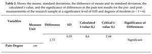
Iraq research ' table

Table 2

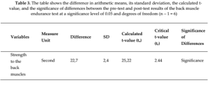
Iraq research ' table

Table 3

Iraq research ' table


1. Lezin N, Watkins-Castillo S. The Hidden Impact of Musculoskeletal Disorders on Americans.

Rosemont, IL; 2018.

Google Scholar

 
2. Clark S, Horton R. Low back pain: a major global challenge. Lancet. 2018;391:2302. doi:10.1016/​S0140-6736(18)30725-6

Google Scholar

 
3. Grabovac I, Dorner TE. Association between low back pain and various everyday performances: Activities of daily living, ability to work and sexual function. Wien Klin Wochenschr. 2019;131:541-549. doi:10.1007/​s00508-019-01542-7

Google Scholar

 
4. Knox J, Orchowski J, Scher DL, et al. The incidence of low back pain in active duty United States military service members. Spine. 2011;36:1492-1500. doi:10.1097/​BRS.0b013e3181f40ddd

Google Scholar

 
5. Elder C, DeBar L, Ritenbaugh C, et al. Comparative effectiveness of usual care with or without chiropractic care in patients with recurrent musculoskeletal back and neck pain. J Gen Intern Med. 2018;33:1469-1477. doi:10.1007/​s11606-018-4539-y

Google Scholar

 
6. Ferreira ML, de Luca K, Haile LM, et al. Global, regional, and national burden of low back pain, 1990–2020, its attributable risk factors, and projections to 2050: a systematic analysis of the Global Burden of Disease Study 2021. Lancet Rheumatol. 2023;5:e316-29. doi:10.1016/​S2665-9913(23)00098-X

Google Scholar

 
7. Carlson H, Carlson N. An overview of the management of persistent musculoskeletal pain. Ther Adv Musculoskelet Dis. 2011;3:91. doi:10.1177/​1759720X11398742

Google Scholar

 
8. Ushida T. Burdensome problems of chronic musculoskeletal pain and future prospects. J Orthop Sci. 2015;20:958. doi:10.1007/​S00776-015-0753-1

Google Scholar

 
9. Inoue S, Kamiya M, Nishihara M, et al. Prevalence, characteristics, and burden of failed back surgery syndrome: the influence of various residual symptoms on patient satisfaction and quality of life as assessed by a nationwide Internet survey in Japan. J Pain Res. 2017;10:811. doi:10.2147/​JPR.S129295

Google Scholar

 
10. Thomson S. Failed back surgery syndrome – definition, epidemiology and demographics. Br J Pain. 2013;7:56. doi:10.1177/​2049463713479096

Google Scholar

 
11. Von Korff MR. Long-term use of opioids for complex chronic pain. Best Pract Res Clin Rheumatol. 2013;27:663-672. doi:10.1016/​j.berh.2013.09.011

Google Scholar

 
12. El-Tallawy SN, Nalamasu R, Salem GI, et al. Management of musculoskeletal pain: an update with emphasis on chronic musculoskeletal pain. Pain Ther. 2021;10:181. doi:10.1007/​S40122-021-00235-2

Google Scholar

 
13. Maher C, Underwood M, Buchbinder R. Non-specific low back pain. Lancet. 2017;389:736-747. doi:10.1016/​S0140-6736(16)30970-9

Google Scholar

 
14. Neprash HT, Barnett ML. Association of primary care clinic appointment time with opioid prescribing. JAMA Netw Open. 2019;2:e1910373. doi:10.1001/​jamanetworkopen.2019.10373

Google Scholar

 
15. Jones CMP, Day RO, Koes BW, et al. Opioid analgesia for acute low back pain and neck pain (the OPAL trial): a randomised placebo-controlled trial. Lancet. Published online July 2023. doi:10.1016/​s0140-6736(23)00404-x

Google Scholar

 
16. Chan CW, Peng P. Failed back surgery syndrome. Pain Med. 2011;12:577-606. doi:10.1111/​j.1526-4637.2011.01089.x

Google Scholar

 
17. Deyo RA, Mirza SK, Turner JA, et al. Overtreating chronic back pain: Time to back off? J Am Board Family Med. 2009;22:62-68.

Google Scholar

 
18. Thomson S, Jacques L. Demographic characteristics of patients with severe neuropathic pain secondary to failed back surgery syndrome. Pain Practice. 2009;9:206-215. doi:10.1111/​j.1533-2500.2009.00276.x

Google Scholar

 
19. Mariconda M, Galasso O, Attingenti P, et al. Frequency and clinical meaning of long-term degenerative changes after lumbar discectomy visualized on imaging tests. European Spine J. 2010;19:136-143. doi:10.1007/​s00586-009-1201-8

Google Scholar

 
20. Keller RB, Atlas SJ, Soule DN, et al. Relationship between rates and outcomes of operative treatment for lumbar disc herniation and spinal stenosis. J Bone Joint Surg. Published online 1999:752-762. doi:10.2106/​00004623-199906000-00002

Google Scholar

 
21. Beckerman D, Esparza M, Lee SI, et al. Cost analysis of single-level lumbar fusions. Global Spine J. 2020;10:39-46. doi:10.1177/​2192568219853251

Google Scholar

 
22. Larson MJ, Adams RS, Ritter GA, et al. Associations of early treatments for low-back pain with military readiness outcomes. J Altern Compl Med. 2018;24:666-676. doi:10.1089/​acm.2017.0290

Google Scholar

 
23. Sherry TB, Roth CP, Bhandarkar M, et al. Chronic Pain Among Service Members: Using Administrative Data to Strengthen Research and Quality Improvement. Published online 2021.

Google Scholar

 
24. Knezevic NN, Mandalia S, Raasch J, et al. Treatment of chronic low back pain – new approaches on the horizon. J Pain Res. 2017;10:1111. doi:10.2147/​JPR.S132769

Google Scholar

 
25. Goertz CM, Long CR, Vining RD, et al. Effect of usual medical care plus chiropractic care vs usual medical care alone on pain and disability among us service members with low back pain: a comparative effectiveness clinical trial. JAMA Netw Open. 2018:1. doi:10.1001/​jamanetworkopen.2018.0105

Google Scholar

 
26. Rubinstein SM, Leboeuf-Yde C, Knol DL, et al. The benefits outweigh the risks for patients undergoing chiropractic care for neck pain: a prospective multicenter, cohort study. J Manipulative Physiol Ther. 2007;30:408-418. doi:10.1016/​j.jmpt.2007.04.013

Google Scholar

 
27. Whedon JM, Toler AWJ, Goehl JM, et al. Association between utilization of chiropractic services for treatment of low-back pain and use of prescription opioids. J Altern Compl Med. 2018;24:552-556. doi:10.1089/​acm.2017.0131

Google Scholar

 
28. Choi J, Lee S, Hwangbo G. Influences of spinal decompression therapy and general traction therapy on the pain, disability, and straight leg raising of patients with intervertebral disc herniation. J Phys Ther Sci. 2015;27:481-483. doi:10.1589/​jpts.27.481

Google Scholar

 
29. Bronfort G, Haas M, Evans RL, et al. Efficacy of spinal manipulation and mobilization for low back pain and neck pain: A systematic review and best evidence synthesis. Spine J. 2004;4:335-356. doi:10.1016/​j.spinee.2003.06.002

Google Scholar

 
30. Whedon JM, Kizhakkeveettil A, Toler AWJ, et al. Initial choice of spinal manipulation reduces escalation of care for chronic low back pain among older medicare beneficiaries. Spine. 2022;47:E142-8. doi:10.1097/​BRS.0000000000004118

Google Scholar

 
31. Coulter ID, Crawford C, Hurwitz EL, et al. Manipulation and mobilization for treating chronic low back pain: a systematic review and meta-analysis. Spine J. 2018;18:866-879. doi:10.1016/​J.SPINEE.2018.01.013

Google Scholar

 
32. Erwin WM. Should you adjust that herniated disc? Thoughts from a chiropractor/molecular scientist. J Can Chiropr Assoc. 2023;67:7.

Google Scholar

 
33. Ohba T, Haro H. TWEAK and TSLP in disc degeneration and spontaneous hernia resorption. JOR Spine. 2020;3. doi:10.1002/​jsp2.1068

Google ScholarPubMed CentralPubMed

 
34. Elkholy AR, Farid AM, Shamhoot EA. Spontaneous resorption of herniated lumbar disk: observational retrospective study in 9 patients. World Neurosurg. 2019;124:e453-9. doi:10.1016/​j.wneu.2018.12.115

Google Scholar

 
35. Yu P, Mao F, Chen J, et al. Characteristics and mechanisms of resorption in lumbar disc herniation. Arthritis Res Ther. 2022:24. doi:10.1186/​s13075-022-02894-8

Google ScholarPubMed CentralPubMed

 
36. Chiu CC, Chuang TY, Chang KH, et al. The probability of spontaneous regression of lumbar herniated disc: a systematic review. Clin Rehabil. 2015;29:184-195. doi:10.1177/​0269215514540919

Google Scholar

 
37. Autio RA, Karppinen J, Niinimäki J, et al. Determinants of spontaneous resorption of intervertebral disc herniations. Spine. 2006;31(11):1247-1252. doi:10.1097/​01.brs.0000217681.83524.4a

Google Scholar

 
38. Takada E, Takahashi M, Shimada K. Natural history of lumbar disc hernia with radicular leg pain: spontaneous MRI changes of the herniated mass and correlation with clinical outcome. J Orthop Surg. 2001;9(1):1-7. doi:10.1177/​230949900100900102

Google Scholar

 
39. Rahme R, Moussa R. The modic vertebral endplate and marrow changes: Pathologic significance and relation to low back pain and segmental instability of the lumbar spine. Am J Neuroradiol. 2008:838-842. doi:10.3174/​ajnr.A0925

Google ScholarPubMed CentralPubMed

 
40. Tu PC, Pan YL, Liang ZQ, et al. Mechanical Stretch Promotes Macrophage Polarization and Inflammation via the RhoA-ROCK/NF- κ B Pathway. Biomed Res Int. 2022;2022. doi:10.1155/​2022/​6871269

Google Scholar

 
41. Cunha C, Silva AJ, Pereira P, et al. The inflammatory response in the regression of lumbar disc herniation. Arthritis Res Ther. 2018;20. doi:10.1186/​s13075-018-1743-4

Google ScholarPubMed CentralPubMed

 
42. Ramos G, Martin W. Effects of vertebral axial decompression on intradiscal pressure. J Neurosurg. 1994;81(3):350-353. doi:10.3171/​jns.1994.81.3.0350

Google Scholar

 
43. Apfel CC, Cakmakkaya OS, Martin W, et al. Restoration of disk height through non-surgical spinal decompression is associated with decreased discogenic low back pain: A retrospective cohort study. BMC Musculoskelet Disord. 2010;11. doi:10.1186/​1471-2474-11-155

Google Scholar


Structural Remodeling of the Lumbar Spine Following Non-Surgical Spinal Decompression: An Automated MRI Study

Please see the full study here.
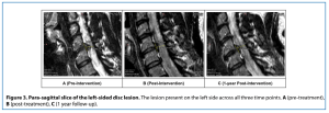
usf cervical study figure

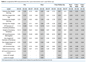
usf cervical study table

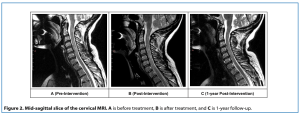
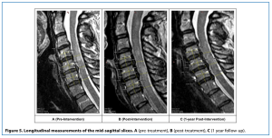
usf cervical study figure mri

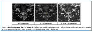
usf cervical study figure mri

usf cervical study figure mri

usf cervical study figure mri

usf cervical study table 

1. Margetis K, Al Khalili Y. Cervical Disc Herniation. Treasure Island, FL: StatPearls Publishing; 2025.

2. Radhakrishnan K, Litchy WJ, O’Fallon WM, Kurland LT. Epidemiology of cervical radiculopathy. Brain 1994;117:325–35.

3. Jordan J, Konstantinou K, O’dowd J. Herniated lumbar disc. BMJ Clin Evid 2009.

4. Epstein NE. A review of complication rates for anterior cervical diskectomy and fusion (ACDF). Surg Neurol Int 2019;10.

5. Yee TJ, Swong K, Park P. Complications of anterior cervical spine surgery: A systematic review of the literature. Journal of Spine Surgery 2020;6:302–22.

6. Chhugani S, Agarwal N, Sheikh F, Borca F, Spanoulis A, Galea I. Cervical spinal degenerative disease in multiple sclerosis. Eur J Neurol 2021;28:2497–502.

7. Xu Q, Tian X, Bao X, Liu D, Zeng F, Sun Q. Nonsurgical spinal decompression system traction combined with electroacupuncture in the treatment of multi-segmental cervical disc herniation: A case report. Medicine (United States) 2022;101:E28540.

8. Ma SY, Kim HD. The effect of motorized spinal decompression delivered via spineMED combined with physical therapy modalities for patients with cervical radiculopathy. J Phys Ther Sci 2010;22:429–35.

9. Schueren S, Luginsland LA, Medina GA, Schilaty ND. Retrospective Chart Review of Nonsurgical Spinal Decompression as a Therapeutic Modality for Low Back Pain. Mil Med 2025;190:134– 40.

10. Connolly JA, Luginsland LA, Welsh S, Bexley JA, Pirone J, Martin WR, et al. Clinical and Imaging Outcomes of Non-Surgical Spinal Decompression for Lumbar Intervertebral Disc Lesions: A Case Series. J Contemporary Chiropr 2025; 8:81–7.

11. Vernon H, Mior S. The Neck Disability Index: a study of reliability and validity. J Manipulative Physiol Ther 1991;14:409–15.

12. Compston A. Aids to the Investigation of Peripheral Nerve Injuries. Medical Research Council: Nerve Injuries Research Committee. His Majesty’s Stationery Office: 1942; pp. 48 (iii) and 74 figures and 7 diagrams; with Aids to the Examination of the Peripheral Nervous System. By Michael O’Brien for the Guarantors of Brain. Saunders Elsevier: 2010; pp. [8] 64 and 94 Figures. Brain 2010;133:2838–44.

13. Ramos G, Martin W. Effects of vertebral axial decompression on intradiscal pressure. J Neurosurg 1994;81:350–3.

14. Roduit N. Weasis DICOM Viewer 2025:Version 4.6.6. Available from: https://github.com/nroduit/Weasis (accessed February 22, 2026)

15. Kuo Y-W, Hsu Y-C, Chuang I-T, Chao P-HG, Wang J-L. Spinal Traction Promotes Molecular Transportation in a Simulated Degenerative Intervertebral Disc Model. Spine (Phila Pa 1976) 2014;39:E550–6.

16. Alrwaily M, Almutiri M, Schneider M. Assessment of variability in traction interventions for patients with low back pain: A systematic review. Chiropr Man Therap 2018;26:1–11.

17. Eldaya RW, Parsons MS, Hutchins TA, Avery R, Burns J, Griffith B, et al. ACR Appropriateness Criteria® Cervical Pain or Cervical Radiculopathy: 2024 Update. J Am Coll Radiol 2025;22:S136–62.

Abstract

Background and Objectives: To investigate MRI changes using SpineReport (r20260928) following non-surgical spinal decompression (NSSD) in patients with chronic low back pain (cLBP). We conducted a retrospective review of patients treated with NSSD who obtained MRI within 6 months of treatment initiation.

Methods: Thirty-six subjects (20 F, 16 M), with a mean age of 55.9 ± 16.4 years, were treated at the USF Health Chiropractic Clinic, Department of Neurosurgery, Brain & Spine. NSSD using DRX9000® (Excite Medical, Tampa, FL, USA) was utilized, with a median of 20 (20, 25) treatments. Treatment levels included L3 (n = 2), L4 (n = 18), and L5 (n = 16). Sagittal T2-weighted images were processed using TotalSpineSeg(r20251124) for automated segmentation, with SpineReport used to quantify disc, vertebrae, spinal canal, and intervertebral foramen metrics.

Results: Mean pain scores improved by 3.4 ± 1.8 from 5.6 ± 1.6 (p < 0.001), while Oswestry Disability Index scores improved by 10.8 ± 9.1 from 17.3 ± 8.0 (p < 0.001). At the treated level, spinal canal cross-sectional area increased from 170.9 to 175.8 mm2 (2.9%, p = 0.040), and right–left diameter increased from 16.0 to 16.3 mm (1.9%, p = 0.025). Disc volume decreased from 7762.8 to 7449.5 mm3 (−4.0%, p = 0.046). No significant changes were observed in disc thickness, DHI, intervertebral foramen area, or vertebral measurements (p > 0.05).

Conclusion: SpineReport enabled automated quantification of key spinal structures. NSSD was associated with increased spinal canal dimensions and improved symptoms. Further research is needed to determine the persistence and clinical significance of these structural changes.

Keywords: non-surgical spinal decompression; magnetic resonance imaging; SpineReport; low back pain

1. Introduction

Low back pain (LBP) is among the most common musculoskeletal disorders worldwide and remains a leading cause of disability, impaired physical function, and reduced quality of life [1,2]. Approximately 80–85% of individuals experience LBP during their lifetime, with nearly one in five developing chronic symptoms (persistent pain >3 months, muscle stiffness, reduced range of motion) [3]. Consequently, LBP places a substantial burden on healthcare systems and society, contributing substantially to the estimated $980 billion spent annually on musculoskeletal disorders in the United States [2,4].
LBP is commonly categorized according to symptom duration as acute (<6 weeks), subacute (6–12 weeks), or chronic (>12 weeks), and by etiology as specific or non-specific [5]. While specific LBP is attributable to an identifiable pathology such as fracture, infection, malignancy, or disc herniation, approximately 80–90% of cases are classified as non-specific because no definitive structural cause can be identified [6,7]. Clinical practice guidelines recommend patient education, exercise therapy, and behavioral interventions as first-line treatment for chronic non-specific LBP [8]. However, many patients continue to experience persistent pain despite conservative care, while alternative treatments such as opioid therapy and spinal fusion are associated with limited long-term benefit, increased complication rates, and substantial healthcare costs [9,10,11,12,13,14,15,16].
These limitations have motivated interest in non-invasive treatment strategies, including non-surgical spinal decompression (NSSD). NSSD uses computer-controlled traction to apply cyclic distraction forces to the lumbar spine with the goal of reducing mechanical loading on the intervertebral discs. The DRX9000®(Excite Medical, Tampa, FL, USA) is one such device developed to treat discogenic LBP. Previous studies have demonstrated improvements in pain and disability following NSSD, accompanied by increases in intervertebral disc height and reductions in disc herniation size [17,18,19,20]. However, the anatomical changes that occur throughout the lumbar spine following treatment remain poorly understood.
Magnetic resonance imaging (MRI) is the preferred imaging modality for evaluating LBP because it provides high-resolution visualization of intervertebral discs, neural structures, paraspinal muscles, and spinal ligaments. Although MRI has become integral to the assessment of spinal pathology, conventional radiologic interpretation remains largely qualitative and is subject to considerable inter-reader variability, potentially influencing diagnosis and treatment decisions [21]. Automated image analysis platforms, such as SpineReport [22], address this limitation by providing standardized, quantitative measurements of spinal anatomy, including disc morphology, spinal canal dimensions, and neural foraminal size. The utility of automated MRI analysis for quantifying anatomical changes following NSSD has not been established.
The purpose of this study was to evaluate whether NSSD treatment is associated with short-term changes in intervertebral disc, spinal canal, neural foramen, and vertebral morphologic metrics in individuals with chronic LBP (cLBP) using automated MRI analysis techniques. We hypothesized that automated MRI analysis would detect measurable changes in spinal canal morphology, intervertebral disc, and neural foraminal anatomy after NSSD treatment.

2. Materials & Methods

This study followed all ethical guidelines for retrospective record review with oversight of the University of South Florida Institutional Review Board (STUDY004354). MRI images were obtained from USF Neurosurgery, Brain & Spine Chiropractic Division (Tampa, FL, USA), Wakeman Chiropractic (Daytona Beach, FL, USA), and Spinal Health & Rehab (Punta Gorda, FL, USA).
Subjects were eligible for analysis if the baseline MRI was obtained within six months before treatment initiation and the follow-up MRI within six months after treatment completion, with no specified maximum interval between the two MRI scans.
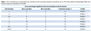
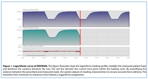
All patients received NSSD treatment with the DRX9000®. Patients were placed in a supine position with the knees flexed to 90°, and a harness was secured around the waist. The initial treatment force was set at 50% of the patient’s body weight (BW) minus 10 lb and progressively increased to 50% BW plus 10 lb. The rate of force progression is individualized for each patient based on comfort.
Additionally, patients completed the numeric pain rating scale (NPRS 0–10; 0 = no pain, 10 = worst imaginable pain) and the Oswestry Disability Index (ODI 0–50; 0 = no disability, 50 = completely disabled) before and after NSSD treatment [23].
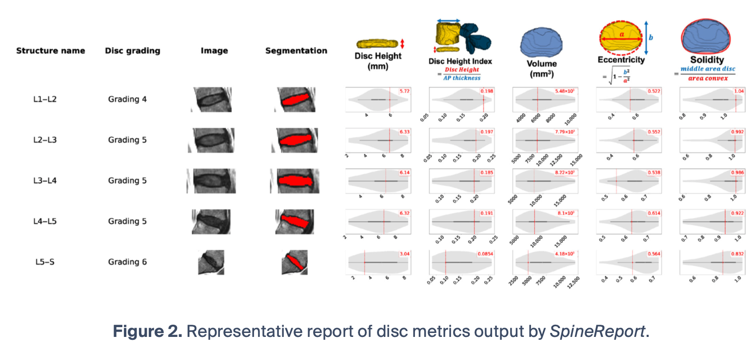
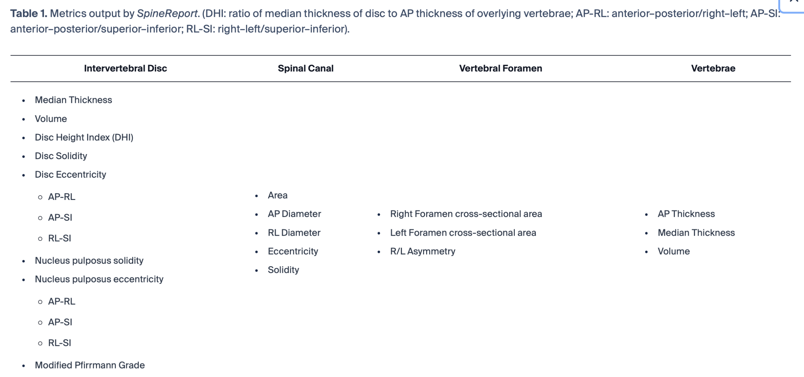
DICOM images were de-identified using a custom Python (v3.10) script and then converted into NIFTI format using the dcm2niix (v1.0.20241211) conversion tool [24]. T2-weighted sagittal images were then processed through TotalSpineSeg (r20251124) [25] to generate segmentations. After segmentation, SpineReport (r20260928) [22] was used to extract the following metrics (Table 1). Spinal canal dimensions were defined for each vertebral level by referencing the inferior border of the disc to the superior border of the overlying vertebral body. These values were then averaged to provide a discrete value for each vertebral level. As the vertebral anatomy was not expected to change during treatment, ICC3,k was calculated for vertebral metrics (AP Diameter, Median Thickness, Volume) to determine stability of vertebral metrics over time. ICC calculations were performed in SPSS (v31) (IBM Corp, Armonk, NY, USA).
usf sep study table

A Mann–Whitney U test was used to determine differences in age, height, and weight between male and female participants at baseline. A matched pairs analysis was performed to elucidate differences between pre- and post-NSSD measures within subjects. A Bowker’s Test was performed for changes in Pfirrmann grading. Additionally, Spearman’s ρ correlation was used to determine the relationship between MRI metrics and patient-reported outcome measures (PROMs). Metrics are reported as change from baseline (Δ = post–pre). Calculations were performed in JMP Pro 19 (SAS Inc, Cary, NC, USA).

3. Results

3.1. Demographics
Sixty-six T2-weighted sagittal MRIs were obtained from the three clinical sites. After applying the inclusion criteria, 30 were excluded due to MRIs being outside the designated window of time. Thus, this study included 36 participants (20 F, 16 M) who underwent both pre- and post-treatment MRI of the lumbar spine. Magnetic field strength ranged from 0.25 to 3.0 T. The median interslice spacing was 5.0 mm (IQR, 4.6–5.0 mm), slice thickness was 4.0 mm (IQR, 4.0–4.0 mm), with a median echo time of 100 ms (IQR, 99–108 ms) and repetition time of 3375 ms (IQR, 2587–3800 ms).
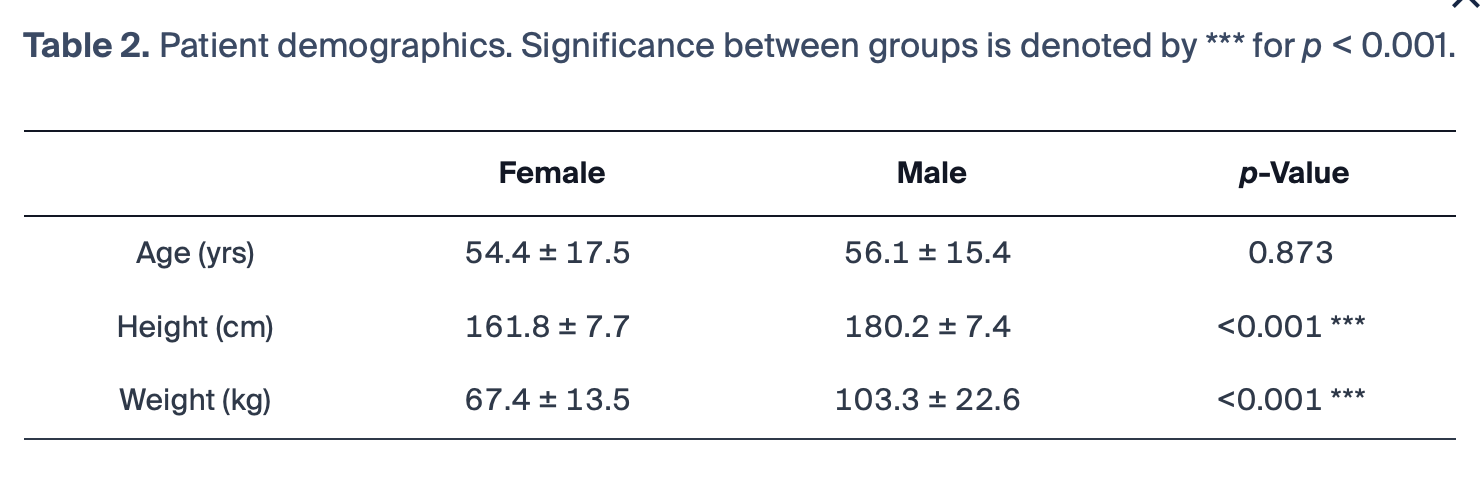
The cohort had a mean age of 55.2 ± 16.4 years, a mean height of 170.0 ± 11.9 cm, and a mean weight of 83.4 ± 25.4 kg. Demographic breakdown by sex is detailed in Table 2. Treatment with the DRX9000®targeted the L3 (n = 2), L4 (n = 18), and L5 (n = 16) levels, with a median of 20 sessions (range 19–29) of 28 min in length. The median interval between MRIs was 141.5 days (range 75–424).
usf sep study table

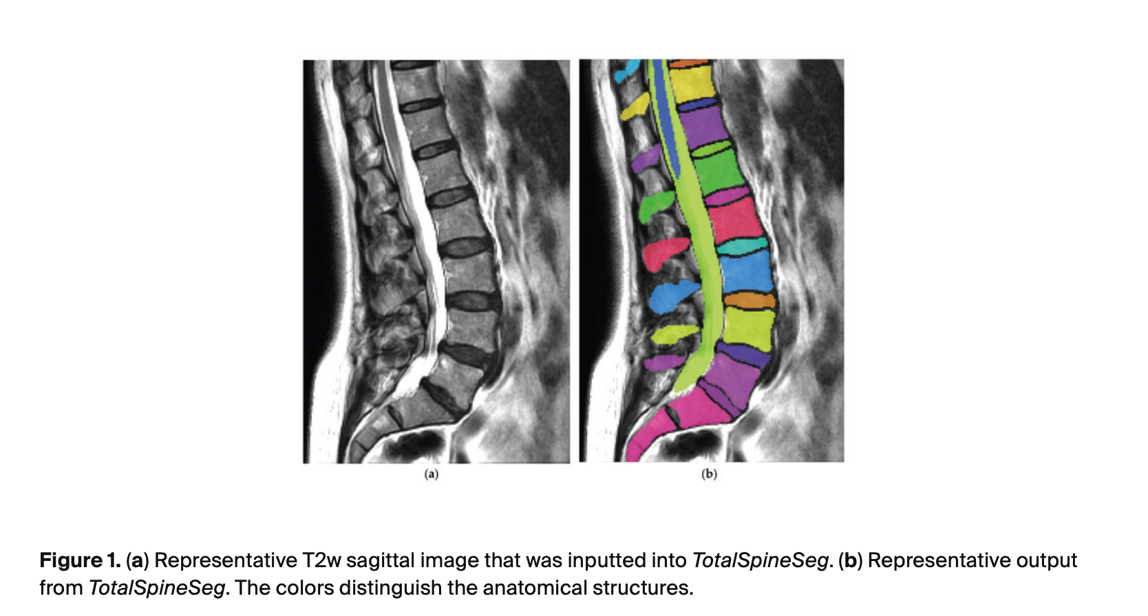
Figure 1a shows an example input image, and Figure 1b shows the TotalSpineSeg output. Figure 2 is a representative report of disc metrics generated by SpineReport.

usf sep study figure

3.2. Post-Treatment

3.2.1. PROMs

Improvements were observed in PROMs following treatment. Mean pain scores improved by 3.4 ± 1.8 points from a baseline of 5.6 ± 1.6 (p < 0.001). Follow-up ODI was available in 20 participants (11 F, 9 M). ODI scores improved by 10.8 ± 9.1 points from a baseline of 17.3 ± 8.0 (p < 0.001).
3.2.2. Disc Measures

At the treated vertebral level, disc volume decreased from 7762.8 mm3 to 7449.6 mm3 (−4.0%, p = 0.046). There were no significant changes in median thickness, DHI, solidity, or eccentricity (p > 0.05). When examining only the subgroup treated at L5, median thickness decreased from 4.4 mm to 3.9 mm (−10.3%, p= 0.009), disc volume decreased from 6223.2 mm3 to 5665.0 mm3 (−9.9%, p = 0.018), and DHI decreased from 0.117 to 0.105 (−11.4%, p = 0.006). No changes were observed in the subgroups treated at L3 or L4.
When correlated with PROMs, there was a correlation between ΔDisc Median Thickness and ΔODI (ρ = −0.4878, p = 0.029). Additionally, in those treated at L4, there was a correlation between ΔNucleus Median Thickness and ΔODI (ρ = −0.7333, p = 0.025).
3.2.3. Pfirrmann Grade

At the level of treatment, five participants showed a reduction in their modified Pfirrmann grade after treatment, two of whom decreased by two levels. Six subjects had an increase in Pfirrmann grade by one level. The remaining 25 showed no change after treatment. The Bowker’s Test revealed no change in Pfirrmann grade among participants (p = 0.996).

3.2.4. Canal Measures

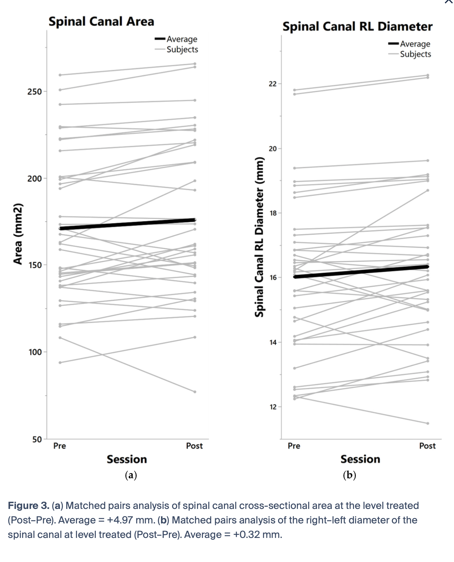
The average area of the spinal canal at the level treated increased from 170.9 mm2 to 175.8 mm2 (2.9%, p = 0.040) (Figure 3a). Right–left (RL) diameter of the canal increased from 16.0 mm to 16.3 mm (1.9%, p = 0.025) (Figure 3b). No changes were seen in the anterior–posterior (AP) diameter of the spinal canal or in spinal canal eccentricity and solidity.
usf sep study figure
3.2.5. Foramen Measures

No changes in foramen area were observed for either the right or left side (p > 0.05) or in RL asymmetry.
3.2.6. Vertebrae Measures

No changes in vertebrae metrics (AP Thickness, Median Thickness, or Volume) were observed after treatment (p > 0.05). AP Thickness had an ICC of 0.983 (0.966, 0.991), Median Thickness = 0.975 (0.950, 0.987), and Volume = 0.996 (0.992 to 0.998).

4. Discussion

The purpose of this study was to evaluate whether NSSD treatment is associated with short-term changes in intervertebral disc, spinal canal, neural foramen, and vertebral morphologic metrics in individuals with cLBP. Following NSSD treatment, participants demonstrated substantial improvements in PROMs, including a mean pain reduction of 3.4 points (60.7%), and a 10.8-point (62.4%) improvement in the ODI. These values exceed the established minimal clinically important differences (MCIDs) for pain (2–2.8 points) and ODI (8.5–10 points) [26,27,28].
These clinical improvements were accompanied by measurable structural changes on MRI. Disc volume decreased by approximately 4%, and within the L5 subgroup, reductions in median disc thickness and disc height index (DHI) were observed, suggesting remodeling of the intervertebral disc. Furthermore, the mean spinal canal cross-sectional area at the treated level increased by 4.9 mm2 (2.9%), while the right–left (RL) canal diameter increased by 0.3 mm (1.9%), indicating increased canal dimensions following treatment. Notably, changes in disc median thickness and nucleus median thickness were strongly correlated with improvements in PROMs, suggesting that alterations in disc morphology may represent one mechanism contributing to improved pain and function after NSSD.
SpineReport has previously demonstrated strong associations between spinal canal anterior–posterior (AP) diameter and cross-sectional area (ρ = 0.59 and 0.56, respectively) and radiologist-graded lumbar central canal stenosis [22]. Additionally, sagittal T1- and T2-weighted MRI scans acquired during the same imaging session demonstrated excellent agreement for disc measurements, with intraclass correlation coefficients (ICCs) ranging from 0.95 to 0.98 for volumetric and thickness-related metrics. Measurements of spinal canal area and AP diameter also showed excellent reproducibility, with ICCs ranging from 0.97 to 0.98. These objective measurements enable reproducible longitudinal assessment of structural changes and have the potential to improve evaluation of treatment response.
Although decreases in disc height can often be considered unfavorable [29,30], the present findings may instead represent adaptive structural remodeling rather than progressive disc degeneration; however, the mechanism underlying the observed reductions in disc volume and thickness remains speculative. One potential explanation is that NSSD treatment alters intradiscal fluid distribution through cyclical loading and unloading of the intended motion segment(s). Previous biomechanical studies have demonstrated that intervertebral discs exhibit dynamic fluid exchange in response to mechanical loading, with changes in hydration status occurring over relatively short time periods [31,32,33,34]. Therefore, reductions in disc dimensions may reflect temporary fluid redistribution rather than structural tissue loss. Alternatively, the observed changes may represent normalization of abnormal disc geometry (e.g., disc herniation) associated with chronic loading and antalgic adaptive patterns that have altered due to improved patient function and decreased LBP. Future studies incorporating quantitative MRI techniques capable of assessing disc hydration and composition will be necessary to better characterize these responses.
In addition to changes in disc morphology, increases in spinal canal area and diameter were observed following treatment. These findings indicate that remodeling of spinal structures may extend beyond the intervertebral disc and may be associated with increased space within the spinal canal. Although the absolute changes in spinal canal dimensions were modest, even small alterations in available neural space may be clinically relevant in individuals with preexisting narrowing from a disc lesion. Because symptoms associated with spinal stenosis and nerve root compression may arise from a combination of mechanical and inflammatory factors, we posit that relatively small anatomical changes could potentially reduce neural irritation and contribute to symptom improvement. While the precise mechanisms underlying these changes remain unclear, they may contribute to the improvements in pain and physical function observed following treatment.
Aside from the changes observed in the disc and spinal canal measures, vertebral measurements served as an internal control, as meaningful changes in vertebral morphology were not expected over the relatively short follow-up period (median 4.7 months). We observed excellent repeatability in vertebrae metrics with ICCs ranging from 0.975 to 0.996. The stability of these vertebral measurements provides validity and increases confidence that the changes observed in disc and canal parameters reflect true anatomical differences rather than variability introduced by imaging acquisition parameters or differences in field of view between imaging sessions.
The improvements observed in PROMs are consistent with previous studies evaluating NSSD treatment [17,20], further supporting the association of NSSD therapy with reductions in pain and disability. To explore the relationship between clinical improvement and structural changes, correlations were examined between changes in PROMs and disc morphology. A significant negative correlation was observed between ΔODI and ΔMedian Disc Thickness and ΔNucleus Median Thickness. Because greater improvements in disability are represented by more negative ΔODI values, this finding indicates that participants who experienced the largest reductions in disability tended to demonstrate increases, or smaller decreases, in median disc thickness following treatment.
Contrary to our hypothesis, neural foraminal dimensions did not demonstrate significant changes following treatment. This may indicate that the observed decompressive effects were localized primarily to the central canal rather than the neural foramina. Alternatively, foraminal changes may be smaller and more variable than changes in central canal dimensions, requiring larger sample sizes to detect reliably. The assessment of these relatively small structures may also have been limited by the resolution of clinical MRI scans, partial volume effects, and variability in imaging hardware and acquisition protocols across sites. Future studies using standardized, higher-resolution imaging and larger cohorts may help determine whether specific patient subgroups exhibit greater foraminal responses.
While current clinical guidelines recommend against routine MRI for patients with acute, subacute, or chronic LBP [35], MRI is still obtained in approximately 16–21% of cases [36]. Given the high lifetime prevalence of LBP, this corresponds to millions of lumbar MRI examinations performed annually. Although MRI plays an important role when clinically indicated, interpretation remains inherently subjective and can vary substantially between radiologists and imaging centers. Herzog et al. demonstrated this variability by sending the same patient to 10 different imaging centers, reporting a global Fleiss’ κ of 0.20 ± 0.06, which indicated poor agreement among radiology reports [21,37]. The authors concluded that the imaging center at which a patient undergoes MRI may directly influence the reported diagnosis, subsequent treatment decisions, and ultimately clinical outcomes. Given the substantial variability in conventional radiology reporting, there is a need for standardized, objective image analysis to complement clinical decision-making. Automated MRI analysis tools such as SpineReport [22] provide reproducible quantitative measurements of clinically relevant spinal morphology while substantially reducing the time required compared with traditional manual measurements.
In the present study, SpineReport detected changes in spinal canal area, spinal canal diameter, and disc morphology following NSSD treatment, whereas vertebral morphologic metrics remained unchanged, supporting the specificity of the observed soft tissue changes. Although structural MRI findings have historically demonstrated weak or inconsistent relationships with pain and disability, significant associations between changes in disc morphology and improvements in ODI scores suggest that quantitative imaging metrics may capture clinically meaningful structural adaptations. While these associations do not establish causality, they support the feasibility of automated quantitative MRI for longitudinal assessment of treatment response and highlight its potential to identify imaging biomarkers that may guide individualized patient management. Future studies with larger cohorts are needed to determine whether these imaging-derived metrics can reliably predict clinical outcomes following NSSD.
Previous investigations of spinal decompression therapies have largely focused on subjective clinical outcomes, whereas objective imaging evidence of structural change remains limited. The present findings therefore contribute important imaging-based evidence demonstrating measurable alterations in spinal canal and disc morphology following treatment. While direct comparison between studies is challenging because of differences in imaging methods, treatment protocols, and patient populations, the current results suggest that structural adaptations may accompany the clinical improvements previously reported with NSSD interventions.

Limitations

Several limitations should be considered when interpreting these findings. This study relied on clinically acquired T2-weighted MRI scans rather than a standardized research imaging protocol. Although imaging data were obtained from multiple radiology centers, all participants included in the final analysis received treatment at a single site (USF Health). Additionally, MRI examinations were performed across different imaging facilities; consequently, acquisition parameters varied between scans, including differences in slice thickness and interslice spacing. The retrospective design precludes causal inference regarding the relationship between NSSD treatment and the observed structural changes, and the absence of a control group makes it difficult to distinguish treatment-related effects from natural variation over time. The relatively modest sample size may have limited statistical power to detect smaller morphological changes and may restrict broader applicability of the findings. While morphologic analyses included 36 participants (20 F, 16 M), follow-up disability outcomes were available for only 20 individuals (11 F, 9 M), further reducing statistical power for analyses involving clinical outcomes. MRI examinations were performed in the supine position, which may not fully reflect spinal morphology under functional loading conditions, but this is a common issue for most clinical scans. Finally, although automated segmentation provides standardized and reproducible measurements, errors related to image quality, segmentation accuracy, and variations in acquisition parameters remain possible.
As this was an exploratory study, additional research is warranted to better characterize the long-term structural effects of NSSD on the intervertebral discs, spinal canal, neural foramina, and vertebral anatomy. Due to the retrospective nature of this study and the lack of a control group, we are unable to determine a causal relationship between NSSD treatment and the improvement in PROMs and MRI metrics we observed. Furthermore, all statistical tests were exploratory, and no corrections for multiple comparisons were applied. Prospective longitudinal studies with larger, multi-center cohorts, standardized imaging protocols, appropriate comparison groups, and comprehensive clinical outcome assessments are needed to confirm these preliminary findings and determine their generalizability across diverse patient populations.

5. Conclusions

Treatment of the lumbar spine with NSSD was associated with increases in spinal canal cross-sectional area and right–left canal diameter, accompanied by a minimal reduction in disc volume. These structural changes occurred alongside improvements in patient-reported pain and disability, suggesting that NSSD may promote short-term remodeling of the intervertebral disc and surrounding spinal anatomy. While the underlying biological mechanisms remain to be fully elucidated, the observed morphological changes may contribute to the clinical improvements reported following treatment. Although the relatively small sample size and heterogeneity in MRI acquisition parameters limit the generalizability of these findings, the results provide convincing preliminary evidence that NSSD may produce clinically meaningful structural and functional changes in individuals with cLBP. Additionally, this study demonstrates the utility of automated, open-source spine analysis tools, including TotalSpineSeg and SpineReport, for objectively quantifying longitudinal changes in spinal morphology, supporting their potential applications in future clinical research and patient monitoring.

Author Contributions

J.A.C.: Conceptualization, Methodology, Validation, Formal Analysis, Investigation, Data Curation, Writing—Original Draft, Visualization. N.M.: Conceptualization, Methodology, Software, Investigation, Writing—Review & Editing. K.A.W.II: Conceptualization, Writing—Review & Editing, Supervision. J.C.-A.: Conceptualization, Software, Writing—Review & Editing, Supervision. N.D.S.: Conceptualization, Methodology, Resources, Writing—Review & Editing, Supervision, Funding acquisition. All authors have read and agreed to the published version of the manuscript.

Funding

Author N.D.S. has received funding from the Florida High-Tech Corridor (FHT 24-19) matched by Excite Medical and the Florida Department of State Center for Neuromusculoskeletal Research.

Institutional Review Board Statement

This study was performed in accordance with the Declaration of Helsinki and all relevant institutional and national guidelines and regulations. The protocol was reviewed and approved for retrospective record review by the University of South Florida Institutional Review Board (STUDY004354; approval date: 8 November 2022).

Data Availability Statement

Data are available upon request.

Acknowledgments

We would like to thank Susan Welsh, Jessie Bexley, and Jamie Pirone from the USF Chiropractic clinic, Peter Wakeman, Josh Hassmann, and Cindy Le from Wakeman Chiropractic, and Kevin Van Nostrand from Spinal Health and Rehab Integrative Medicine for providing us with the MRIs needed for this study. We would also like to acknowledge Excite Medical, the Florida Department of State Center for Neuromusculoskeletal Research, and the Florida High-Tech Corridor for funding this work.

Conflicts of Interest

Author N.D.S. has received Florida High-Tech Corridor funding in partnership with Excite Medical, the manufacturer of the DRX9000®. Excite Medical recognizes that under University policy the results of sponsored research must be publishable and agrees that the University Project Director or other University employees engaged in the project are permitted to present at symposia and professional meetings and to publish in journals, theses or dissertations, or otherwise of their own choosing, the data, methods, and results of the project either positive or negative. All other authors declare no competing interests.

References

  1. Clark, S.; Horton, R. Low back pain: A major global challenge. Lancet 2018, 391, 2302. [Google Scholar] [CrossRef] [Scilit] [PubMed]
  2. Lezin, N.; Watkins-Castillo, S. The Hidden Impact of Musculoskeletal Disorders on Americans; United States Bone and Joint Initiative: Rosemont, IL, USA, 2018. [Google Scholar]
  3. Grabovac, I.; Dorner, T.E. Association between low back pain and various everyday performances: Activities of daily living, ability to work and sexual function. Wien. Klin. Wochenschr. 2019, 131, 541–549. [Google Scholar] [PubMed]
  4. Elder, C.; DeBar, L.; Ritenbaugh, C.; Dickerson, J.; Vollmer, W.M.; Deyo, R.A.; Johnson, E.S.; Haas, M. Comparative Effectiveness of Usual Care With or Without Chiropractic Care in Patients with Recurrent Musculoskeletal Back and Neck Pain. J. Gen. Intern. Med. 2018, 33, 1469–1477. [Google Scholar] [CrossRef] [Scilit] [PubMed]
  5. Furlan, A.D.; Malmivaara, A.; Chou, R.; Maher, C.G.; Deyo, R.A.; Schoene, M.; Van Tulder, M.W. 2015 Updated Method Guideline for Systematic Reviews in the Cochrane Back and Neck Group. Spine 2015, 40, 1660–1673. [Google Scholar] [CrossRef] [Scilit] [PubMed]
  6. Hartvigsen, J.; Hancock, M.J.; Kongsted, A.; Louw, Q.; Ferreira, M.L.; Genevay, S.; Hoy, D.; Karppinen, J.; Pransky, G.; Sieper, J.; et al. What low back pain is and why we need to pay attention. Lancet 2018, 391, 2356–2367. [Google Scholar] [CrossRef] [Scilit] [PubMed]
  7. Koes, B.; Herbert, R.; Maher, C. High-force traction did not improve the clinical course of sub-acute and chronic non-specific low back pain. Aust. J. Physiother. 1998, 44, 213–214. [Google Scholar]
  8. Chiarotto, A.; Koes, B.W. Nonspecific Low Back Pain. N. Engl. J. Med. 2022, 386, 1732–1740. [Google Scholar] [CrossRef] [Scilit] [PubMed]
  9. Beckerman, D.; Esparza, M.; Lee, S.I.; Berven, S.H.; Bederman, S.S.; Hu, S.S.; Burch, S.; Deviren, V.; Tay, B.; Mummaneni, P.V.; et al. Cost Analysis of Single-Level Lumbar Fusions. Glob. Spine J. 2019, 10, 39–46. [Google Scholar] [CrossRef] [Scilit] [PubMed]
  10. Hayden, J.A.; Ellis, J.; Ogilvie, R.; Malmivaara, A.; van Tulder, M.W. Exercise therapy for chronic low back pain. Cochrane Database Syst. Rev. 2021, 9, CD009790. [Google Scholar] [CrossRef] [Scilit] [PubMed]
  11. Henschke, N.; Ostelo, R.W.; Van Tulder, M.W.; Vlaeyen, J.W.; Morley, S.; Assendelft, W.J.; Main, C.J. Behavioural treatment for chronic low-back pain. Cochrane Database Syst. Rev. 2010, 2011, CD002014. [Google Scholar] [CrossRef] [Scilit] [PubMed]
  12. Inoue, S.; Kamiya, M.; Nishihara, M.; Arai, Y.-C.P.; Ikemoto, T.; Ushida, T. Prevalence, characteristics, and burden of failed back surgery syndrome: The influence of various residual symptoms on patient satisfaction and quality of life as assessed by a nationwide Internet survey in Japan. J. Pain Res. 2017, ume 10, 811–823. [Google Scholar] [CrossRef] [Scilit] [PubMed]
  13. Jones, C.M.P.; Day, R.O.; Koes, B.W.; Latimer, J.; Maher, C.G.; McLachlan, A.J.; Billot, L.; Shan, S.; Lin, C.-W.C.; McLachlan, H.; et al. Opioid analgesia for acute low back pain and neck pain (the OPAL trial): A randomised placebo-controlled trial. Lancet 2023, 402, 304–312, Correction in Lancet 2023, 402, 10402, https://doi.org/10.1016/S0140-6736(23)01681-1. [Google Scholar] [CrossRef] [Scilit] [PubMed]
  14. Maher, C.; Underwood, M.; Buchbinder, R. Non-specific low back pain. Lancet 2017, 389, 736–747. [Google Scholar] [CrossRef] [Scilit] [PubMed]
  15. Moghib, K.; Altalab, G.; Jader, A.; Ghanm, T.I.E.; Hijazy, M.; Tarawneh, D.Y.; Hannat, R.; Salomon, I.; Edress, A.I.; Abu Arafeh, M.W.; et al. Comparison between spinal fusion vs. nonoperative treatment for lumbar degenerative pathology: A systematic review and meta-analysis. Neurosurg. Rev. 2025, 48, 502. [Google Scholar] [CrossRef] [Scilit] [PubMed]
  16. Zaina, F.; Tomkins-Lane, C.; Carragee, E.; Negrini, S. Surgical versus non-surgical treatment for lumbar spinal stenosis. Cochrane Database Syst. Rev. 2016, 2016, CD010264. [Google Scholar] [CrossRef] [Scilit] [PubMed]
  17. Connolly, J.; Luginsland, L.; Welsh, S.; Bexley, J.A.; Pirone, J.; Martin, W.; Thorpe, S.; Schilaty, N.D. Clinical and Imaging Outcomes of Non-Surgical Spinal Decompression for Lumbar Intervertebral Disc Lesions: A Case Series. J. Contemp. Chiropr. 2025, 8, 81–87. [Google Scholar]
  18. Connolly, J.A.; Gabro, A.M.; Welsh, S.; Bexley, J.; Schilaty, N.D. Cervical Non-Surgical Spinal Decompression for Disc Herniation in a Patient with Multiple Sclerosis: A Case Study Using the DRX9000C. J. Phys. Med. Rehabil. 2026, 8,

The DRX9000® is the Worlds Most Advanced Non-Surgical Treatment for Back and Neck Pain